Anatomy Chest Muscles Diagram / Muscles Of The Trunk Anatomy Diagram Pictures Kenhub : All about the chest muscles.. In this article, we shall learn about the anatomy of the muscles of the anterior chest. When you are taking anatomy and physiology you will be required to identify major muscles in the human body. Attached to the bones of the skeletal system are about 700 named muscles that make up roughly half of a person's body weight. Human anatomy and physiology diagrams: Find out more about the individual muscles within the chest anatomy by clicking their respective links throughout this page.
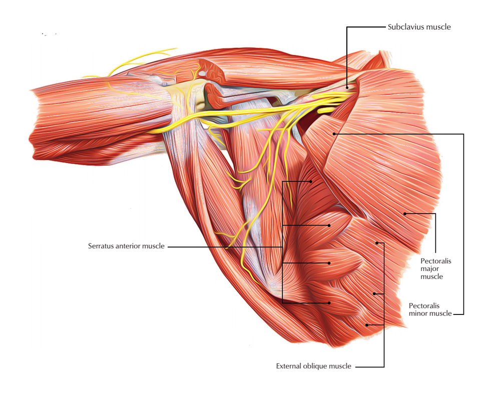
The pectoralis minor muscle (not shown in the diagram) is located underneath the pectoralis major muscle, attaching to the coracoid. Find out more about the individual muscles within the chest anatomy by clicking their respective links throughout this page. If you need to review the human skeleton for an upcoming test or quiz, this page provides several free human skeleton diagrams to help you study. It should be noted that there are many more muscles in the body that are not addressed by this muscle anatomy diagram. Almost every movement in the body is the outcome of muscle contraction.
Easy Notes On The Pectoral Region Muscles Learn In Just 6 Mins Earth S Lab from www.earthslab.com Related posts of chest muscles diagram. The pectoralis minor muscle (not shown in the diagram) is located underneath the pectoralis major muscle, attaching to the coracoid. Muscle anatomy anterior muscular anatomy for pilates on pinterest grays anatomy muscle, picture of muscle anatomy anterior muscular anatomy for pilates rehabilitating acute hamstring injuries | el paso, tx chiropractor. The gastrocnemius and soleus muscles taper and merge at the base of the calf muscle. Human anatomy and physiology diagrams: Their main function is contractibility. Muscles, connected to bones or internal organs and blood vessels, are in charge for movement. The first is the pectoralis major which is the largest one and located in the center of the chest.
Related posts of chest muscles diagram.
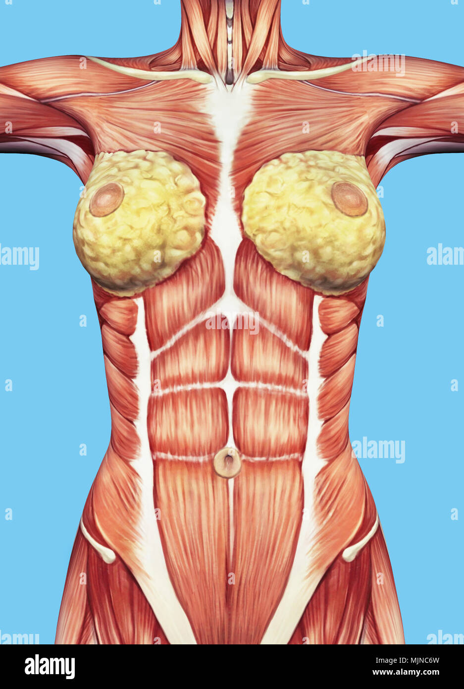
Anatomy • free medical books. Related posts of chest muscles diagram. Find out more about the individual muscles within the chest anatomy by clicking their respective links throughout this page. Almost every movement in the body is the outcome of muscle contraction. The pectorals, or chest muscles, are so large and prominent that they can't be hidden. Human anatomy and physiology diagrams: Adducts & flexes the arm (humerus). In this article, we shall learn about the anatomy of the muscles of the anterior chest. Female chest muscle anatomy diagram ~ diagram. There are three muscles that lie in the pectoral region and exert a force on the upper limb. Meet your pectoralis major and pectoralis minor. Chest anatomy muscles human chest muscle anatomy diagram chest muscles anatomy picture female chest muscle anatomy diagram. We think this is the most useful anatomy picture that.
They are the pectoralis major, pectoralis minor, and the serratus anterior. This page provides an overview of the chest muscle group. Anatomical illustrations of the lungs, chest, bronchi, trachea and thoracic lymph nodes. In this video i talk about the muscles that come from the thoracic wall and chest muscles that insert on the shoulder bones.✅. The gastrocnemius and soleus muscles taper and merge at the base of the calf muscle.
Paint Draw Paint Learn To Draw Anatomy Basics The Chest Muscle from 4.bp.blogspot.com Because of its size it is the strongest one and mainly it is responsible for having huge chest. A massive chest anchors the upper body and enhances the appearance of your shoulders, arms, and abs. Female chest muscle anatomy diagram ~ diagram. The chest anatomy includes the pectoralis major, pectoralis minor and the serratus anterior. This muscle moves each shoulder joint in four distinct ways as well as keeps the arms attached to the body. They are the pectoralis major, pectoralis minor, and the serratus anterior. The pectoralis major muscles (also known as the pecs) are located on the front of the rib cage, and form the major muscles of the chest. Tough connective tissue at the bottom of the calf muscle merges with the achilles tendon.
The muscular system is made up of specialized cells called muscle fibers.
Adducts & flexes the arm (humerus). This page provides an overview of the chest muscle group. Start studying chest muscles anatomy. In this article, we shall learn about the anatomy of the muscles of the anterior chest. 1024 x 873 jpeg 135 кб. Because of its size it is the strongest one and mainly it is responsible for having huge chest. Located in the rib cage, this muscle keeps the shoulder blade against the chest wall and helps rotate the shoulder blade upward. Identify the muscle labeled as 1 in the diagram above In this image, you will find part of the pectoral muscles mainly used in it. Meet your pectoralis major and pectoralis minor. Human anatomy diagram shoulder anatomy shoulder muscles shoulder muscles and chest. Their main function is contractibility. Find out more about the individual muscles within the chest anatomy by clicking their respective links throughout this page.
Located immediately below the skin) muscles of the body. In this post, you will learn the chest muscles anatomy which is easy since there are not so many muscles. At the level of the pelvic bones the abdomen ends and the pelvis begins. We think this is the most useful anatomy picture that. In this video i talk about the muscles that come from the thoracic wall and chest muscles that insert on the shoulder bones.✅.
Chest Muscle Anatomy Anatomy Drawing Diagram from c8.alamy.com In this image, you will find part of the pectoral muscles mainly used in it. Chest muscles anatomy for bodybuilders. The gastrocnemius and soleus muscles taper and merge at the base of the calf muscle. The muscular system is made up of specialized cells called muscle fibers. If you need to review the human skeleton for an upcoming test or quiz, this page provides several free human skeleton diagrams to help you study. Identify the muscle labeled as 1 in the diagram above Start studying chest muscles anatomy. Attached to the bones of the skeletal system are about 700 named muscles that make up roughly half of a person's body weight.
The interactive muscle anatomy diagram shown below outlines the major superficial (i.e.
All about the chest muscles. The pectorals, or chest muscles, are so large and prominent that they can't be hidden. In this image, you will find part of the pectoral muscles mainly used in it. The gastrocnemius and soleus muscles taper and merge at the base of the calf muscle. Muscle anatomy quiz for anatomy and physiology! Find out more about the individual muscles within the chest anatomy by clicking their respective links throughout this page. Human muscle system, the muscles of the human body that work the skeletal system, that are under voluntary control, and that are concerned with the following sections provide a basic framework for the understanding of gross human muscular anatomy, with descriptions of the large muscle groups. The first is the pectoralis major which is the largest one and located in the center of the chest. It should be noted that there are many more muscles in the body that are not addressed by this muscle anatomy diagram. A massive chest anchors the upper body and enhances the appearance of your shoulders, arms, and abs. Almost every movement in the body is the outcome of muscle contraction. The pectoralis minor muscle (not shown in the diagram) is located underneath the pectoralis major muscle, attaching to the coracoid. Muscle anatomy anterior muscular anatomy for pilates on pinterest grays anatomy muscle, picture of muscle anatomy anterior muscular anatomy for pilates rehabilitating acute hamstring injuries | el paso, tx chiropractor.
There are three muscles that lie in the pectoral region and exert a force on the upper limb chest muscles diagram. Tough connective tissue at the bottom of the calf muscle merges with the achilles tendon.


0 Komentar